OncoBLAZE

TM

THE CHALLENGE
Cancer Recurrence Remains a Major Challenge

20M+
New cancer cases globally each year


2M+
New cancer cases in the United States annually

Residual Cells
Remain after surgery, up to 40% in some cancer types.


Recurrence
Leads to additional, costly treatments and poorer outcome.

While surgery is often highly effective, microscopic cancer cells can remain behind after tumor removal. These residual cells may eventually grow and lead to recurrence, requiring additional treatment and increasing the burden on patients and healthcare systems.
OUR VISION
Building the Future of Image-Guided Cancer Therapy
OncoBLAZE is developing technologies that enable precise chemo drug delivery and real-time visualization of therapy. Our goal is to improve outcomes while reducing unnecessary exposure to healthy tissue.

Triggered Nanoparticles
Safely transport chemotherapy through the body while preventing unwanted drug release.


Precision Energy Delivery
Our proprietary device delivers targeted electromagnetic energy to the surgical cavity with high spatial accuracy.

Visualization of Chemo Drug Delivery
Our proprietary imaging technology enables visualization of drug release and treatment response during therapy.

HOW ONCOBLAZE WORKS
Built to Target What Surgery Leaves Behind
Traditional chemotherapy distributes throughout the body, exposing healthy tissues to toxic drugs while delivering only a small fraction to cancer cells.
OncoBLAZE takes a different approach.
Step 1:
Nanoparticle Administration
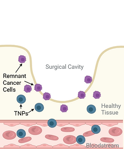
Step 2:
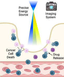
Targeted Drug Release
Step 3:
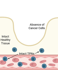
Complete Eradication
Tumor is surgically removed, leaving a surgical cavity that may contain residual cancer cells. TNP containing chemotherapy are administered intravenously, circulating throughout the body without releasing the drug. Release is activated locally in the cavity by targeted, mild heat.
Energy is directed precisely to the remnant cancer cells using the imaging-guided system. The heat triggers TNP rupture, causing the nanoparticles to release chemotherapy only where treatment is needed and inducing cancer cell death.
Residual cancer cells are eliminated while minimizing exposure to healthy tissue, resulting in a clean surgical cavity bed with intact surrounding tissue and no remaining cancer cells.
PROPRIETARY IMAGING TECHNOLOGY
See Drug Delivery in Real Time
We developed imaging technology that visualizes drug released from TNP during heating. This enables the physician to deliver drug precisely to the tissue where remnant cancer cells are, and to ensure adequate drug is delivered to kill all cancer cells.
Heat is precisely targeted to a portion of the surgical cavity where a tumor has been removed, and where remnant cancer cells are anticipated. The amount of drug delivered to the heated tissue is visualized while heat is applied.
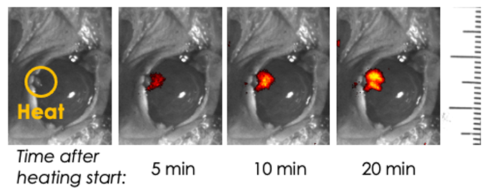
Real-time visualization of drug release during targeted heating.
No enclosed chamber.No interruption of the experiment.
Researchers can inject, dose, heat, operate, and access the animal while continuously visualizing fluorescence data in real time with high spatial resolution.
COMING SOON
SphynxVision™
OncoBLAZE has developed a new preclinical imaging platform based on the proprietary imaging technology designed for clinical use.
Real-time fluorescence imaging
under ambient light with an open
workflow and high spatial resolution.
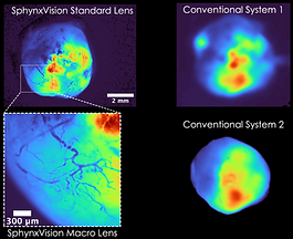
GFP-labeled tumor imaged ex vivo. SphynxVision reveals tumor-associated microvasculature (<100 µm vessel diameter) while maintaining an open imaging workflow.


-
High-resolution imaging reveals fine vascular and tissue structures
-
Sensitivity comparable to leading commercial imaging platforms
-
Multi-wavelength imaging from visible to NIR spectra
-
Dynamic imaging captures biological processes in real time
-
Continuous access enables intervention without interrupting imaging